
Heart and vascular diagnostics · Saint Petersburg
Intravascular Coronary Diagnostics: FFR, IVUS and OCT
Measuring fractional flow reserve and looking at the artery from the inside during coronary angiography. They help decide precisely whether a stent is needed and check how it was placed.
Paid or under CHI
free with a referral and indications
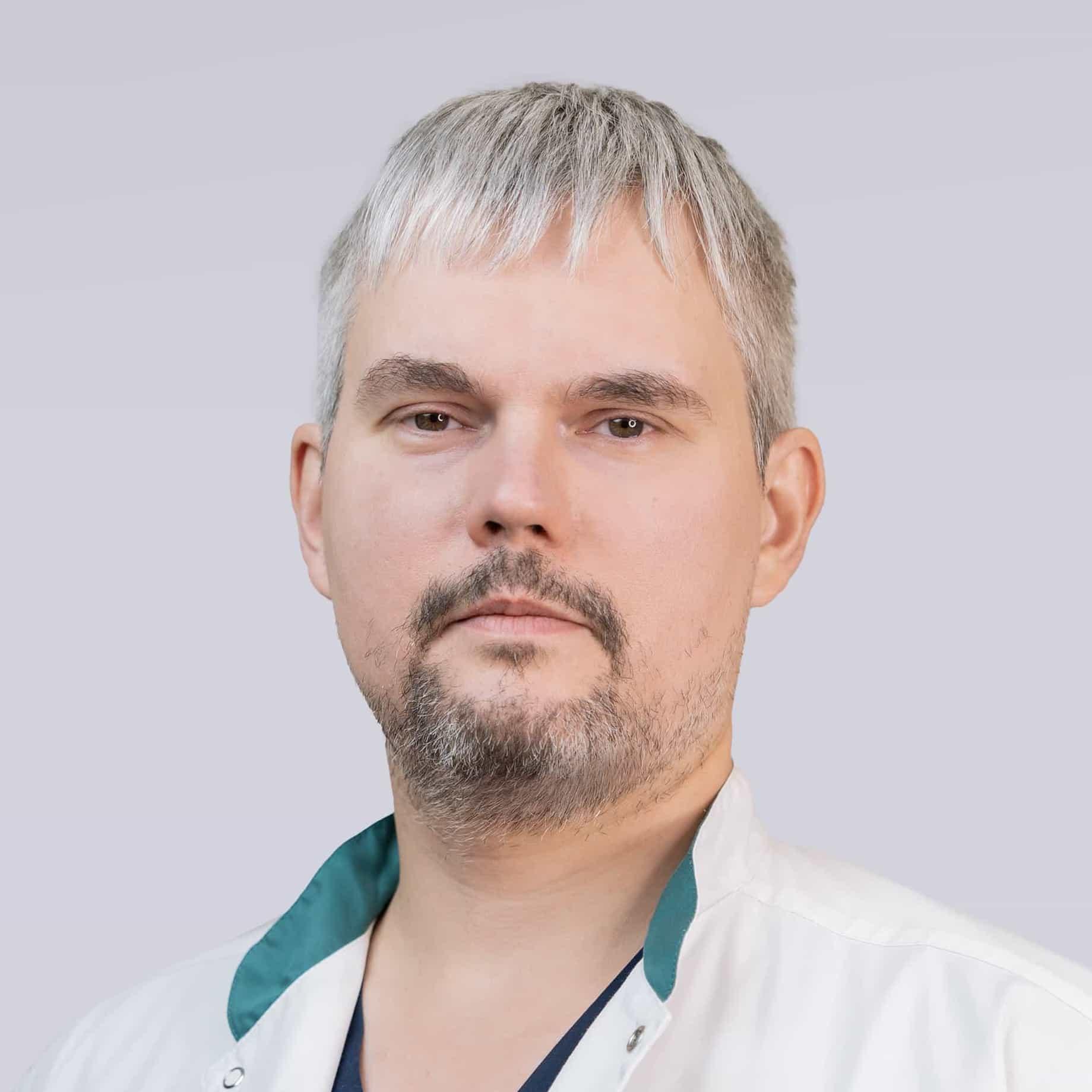 Text reviewed by Alexey Vasilev, endovascular surgeon
What is intravascular diagnostics
Intravascular diagnostics is a group of methods that complement coronary angiography. Angiography shows the silhouette of the artery's lumen filled with contrast — and nothing more. From that silhouette it is not always clear whether a narrowing limits blood flow or what the plaque causing it is like. Intravascular methods answer these questions.
FFR (fractional flow reserve) and iFR (instantaneous wave-free ratio) measure the pressure in the artery before and after a narrowing with a thin pressure wire — to find out whether the stenosis causes ischemia. IVUS (intravascular ultrasound) and OCT (optical coherence tomography) show the artery wall from the inside, as a cross-section, using ultrasound or infrared light.
These methods matter most for «borderline» narrowings of 50–90 %, whose significance cannot be judged by eye. Large randomised trials have shown that deciding on stenting by FFR reduces unnecessary stents and improves outcomes, and that stent placement guided by IVUS or OCT lowers the risk of repeat procedures. The studies are done through the same access as the angiography, under local anesthesia — no separate procedure is needed.
Intravascular diagnostics in numbers
FFR, IVUS and OCT are performed in the cath lab during coronary angiography or stenting. The patient is awake, with ECG and blood pressure monitored.
- added to coronary angiography
- +10–20 min
- FFR threshold: below it a stenosis is significant
- 0.80
- iFR threshold, no drugs needed
- 0.89
- OCT resolution — 10 times finer than IVUS
- 10–20 µm
When FFR, IVUS and OCT are needed
An interventional cardiologist decides on an intravascular study during coronary angiography or in advance, when planning a procedure:
- A coronary narrowing of 50–90 % without proven ischemia on stress tests
- Multivessel disease — finding which narrowings really need treatment
- Choosing between stenting and coronary artery bypass grafting
- Left main coronary artery disease — assessing the lumen area
- Long, calcified or bifurcation lesions — planning stent size and vessel preparation
- Checking the result of stenting: stent expansion, apposition to the wall, edges
- Restenosis or thrombosis of an earlier stent — finding the cause
- Acute coronary syndrome of unclear cause — plaque rupture, erosion, arterial dissection
Limitations
- Contraindications are the same as for coronary angiography: severe general condition, contrast intolerance, acute kidney failure
- For FFR with ATP or adenosine — severe bronchial asthma, high-grade atrioventricular block; in these cases drug-free iFR is used
- OCT needs extra contrast, so IVUS is preferred when kidney function is reduced
What each method shows
The methods answer different questions and are often used together:
FFR — is the narrowing significant
The ratio of pressure beyond the narrowing to aortic pressure at maximum flow. FFR of 0.80 or less means the stenosis causes ischemia and restoring flow will help; above 0.80 the narrowing can safely be treated with drugs.
iFR — the same without drugs
The instantaneous wave-free ratio is measured while the heart relaxes, without ATP or adenosine. A value of 0.89 or less is considered significant.
IVUS — size and plaque
The true artery diameter, plaque volume, lumen area, calcium. It penetrates deep into the wall, so it is indispensable for left main disease and stent sizing.
OCT — surface detail
Images at micron resolution: the thin cap of a vulnerable plaque, rupture, thrombus, calcium thickness, every stent strut and its apposition to the wall.
Choosing the stent
From intravascular images the surgeon chooses the stent's diameter and length and where its edges will land on healthy wall, and, with heavy calcification, how to prepare the vessel.
Checking the result
After stenting — is the stent fully expanded, does it sit tight against the wall, are there tears at the edges. Shortcomings are fixed at once, in the same procedure.
FFR, IVUS and OCT are invasive: they are done only inside the artery during coronary angiography or stenting. Stress tests (stress echocardiography, treadmill test) and CT coronary angiography do not replace them, but help to tell in advance whether invasive diagnostics is needed.
Preparation
Intravascular studies need no separate preparation — it is the same as for coronary angiography:
- Complete blood count and biochemistry (including creatinine), coagulation tests, infection tests, an ECG and echocardiography
- On the day of the procedure — no food; water and essential medicines as the doctor advises
- Discuss anticoagulants, metformin and diuretics with the doctor in advance
- Avoid coffee, strong tea and chocolate for 12–24 hours: caffeine weakens the drugs given for FFR measurement
- Report any allergy to iodine or contrast agents, bronchial asthma or kidney disease
- Bring stress test results, a previous angiography (disc with the recording) and discharge summaries
How the study is done
Intravascular diagnostics is performed by an interventional cardiologist in the cath lab. It adds 10–20 minutes to coronary angiography.
Coronary angiography
Under local anesthesia a catheter is passed through an artery in the wrist (less often the groin) to the coronary arteries, and coronary angiography is performed.
Passing the sensor
If a narrowing of uncertain significance is found, a thin pressure wire or an IVUS / OCT catheter is passed through the catheter beyond the narrowing. The patient does not feel this.
Measuring FFR
For FFR a vasodilator (ATP or adenosine) is given. For 1–2 minutes you may feel warmth, chest tightness or shortness of breath — the expected and short-lived effect of the drug. iFR needs no drug.
Recording images
The IVUS or OCT catheter is pulled back automatically along the artery, recording images of its wall. For OCT the artery is flushed with contrast for 2–3 seconds during recording.
Decision on the spot
Based on the results the surgeon decides at once whether stenting is needed and, if so, performs it in the same procedure. In complex multivessel disease the decision on bypass surgery is made by a heart team of cardiac surgeons and cardiologists.
Understanding the results
FFR and iFR results are numbers. FFR above 0.80 (iFR above 0.89) means the narrowing does not limit flow enough to cause ischemia: such a stenosis is safe to treat with drugs, and a stent will not improve the prognosis. FFR of 0.80 or less means the narrowing is significant and restoring flow will help.
IVUS and OCT measure the minimum lumen area and the size of the artery and plaque. For the left main coronary artery an IVUS lumen area below about 6 mm² is considered significant. After stenting, the lumen area inside the stent is compared with healthy segments of the artery.
The results go into the angiography report, and the surgeon explains them to the patient on the day of the procedure. A normal FFR does not mean there is no atherosclerosis: atherosclerosis remains, and the treatment prescribed by the cardiologist must continue — statins, blood pressure and sugar control, quitting smoking.
If you have already had a coronary angiography and were advised a stent or bypass surgery but have doubts, you can get a consultation at the Center by sending the disc with the recording and your discharge summaries.
FFR, IVUS or OCT: the difference
The methods do not compete but complement each other. This is how they are chosen:
Angiography with and without FFR
Angiography shows how narrow the lumen is, but not whether the narrowing causes ischemia. A 70 % narrowing may be insignificant and a 50 % one significant. FFR gives a precise answer for a particular stenosis.
FFR and iFR
Both measure pressure in the artery. FFR needs a drug, iFR does not, so measurement is quicker and suits people with asthma. Treatment outcomes with the two are comparable.
IVUS and OCT
IVUS penetrates deeper into the wall and better shows the true artery size and the left main, with no extra contrast. OCT gives a 10 times sharper image of the surface: vulnerable plaques, clots, stent apposition.
Invasive and non-invasive tests
Stress echo and CT coronary angiography estimate the likelihood of a significant narrowing without entering the body. FFR is the reference standard, done when the question must be settled right before stenting.
FFR, IVUS and OCT price in Saint Petersburg
- Coronary angiographyIncludes all consumables and observation
- 57,600 ₽
- Additional studies (FFR, OCT)When clinically indicated
- 97,600 ₽
The final price depends on the method and the number of arteries examined — the doctor will name it after reviewing your documents. Intravascular diagnostics is free under CHI and a federal quota when it is part of the treatment.
Paid or under CHI
When indicated, FFR, IVUS and OCT are done free of charge — as part of coronary angiography and stenting under Compulsory Health Insurance (CHI) and federal quotas for high-tech medical care. To get treatment, send your documents to the Center: a cardiac surgeon will review them and give a decision.
FFR, IVUS and OCT questions
FFR (fractional flow reserve) shows what share of normal blood flow a narrowed artery lets through. It is measured with a thin pressure wire during coronary angiography. If FFR is 0.80 or less, the narrowing impairs the heart's blood supply and is worth treating; if higher, drugs are enough.
Not always. The percentage on an angiogram is an estimate by eye, and a 70 % stenosis may not cause ischemia while a 50 % one may. The decision takes into account symptoms, stress test results and, when in doubt, FFR or iFR measurement.
No pain: the procedure is done under local anesthesia. When the FFR drug is given you may feel warmth and chest tightness for 1–2 minutes — an expected effect that passes quickly. The risk is comparable to that of coronary angiography itself.
Both show the artery wall from the inside. IVUS uses ultrasound: it «sees» deeper and is better for assessing artery size and the left main. OCT uses light: the image is 10 times sharper and shows fine details of plaque and stent, but recording needs extra contrast.
Invasive FFR is measured only inside a coronary artery, that is, during coronary angiography or stenting. The likelihood of a significant narrowing is estimated non-invasively with stress echocardiography and CT coronary angiography.
Usually 10–20 minutes to the coronary angiography. Observation afterwards is the same as after a standard angiography.
No, preparation is the same as for coronary angiography. In addition, avoid coffee, strong tea and chocolate for 12–24 hours: caffeine weakens the drugs used for FFR.
A normal FFR means that particular narrowing does not limit blood flow now and a stent is not needed. But atherosclerosis does not go away: keep up the treatment prescribed by the cardiologist and control your risk factors.
Yes. When indicated, these studies are done as part of coronary angiography and stenting under CHI and federal quotas. Paid additional studies (FFR, OCT) cost 97,600 ₽; the doctor will name the final price.
Conditions it helps to diagnose
- AtherosclerosisNarrowing of arteries due to cholesterol plaques, leading to impaired blood flow and risk of heart attack, stroke, or gangrene.Learn more
- Ischemic Heart DiseaseNarrowing of the heart vessels, causing angina, heart attack, or death.Learn more
- Ischemic CardiomyopathyImpaired blood supply to the heart, causing its enlargement and heart failure.Learn more
Related procedures and diagnostics
- CoronarographyAn accurate method of diagnosing the condition of the coronary arteries of the heart for the detection and treatment of diseases.Learn more
- Coronary artery stentingMinimally invasive surgery that restores blood flow in the arteries of the heart, preventing heart attacks and other complications.Learn more
- Coronary artery bypass graftingRestores blood flow to the heart, bypassing narrowed arteries, improving the condition of coronary heart disease.Learn more
How to get care at the Center
- See a cardiologistConsultation and tests at the Outpatient Cardiology Department on Stachek Square.Learn more
- Free under CHI and quotasStep-by-step guide: which documents you need and how to get treatment free of charge.Learn more
- Online expert opinionAn online talk with a cardiologist about your documents — for patients from other cities.Learn more
Still have questions?
Book an online talk or get in touch with our administrators. We'll help you choose the right specialist, answer any organizational questions, and explain how to prepare for the consultation.